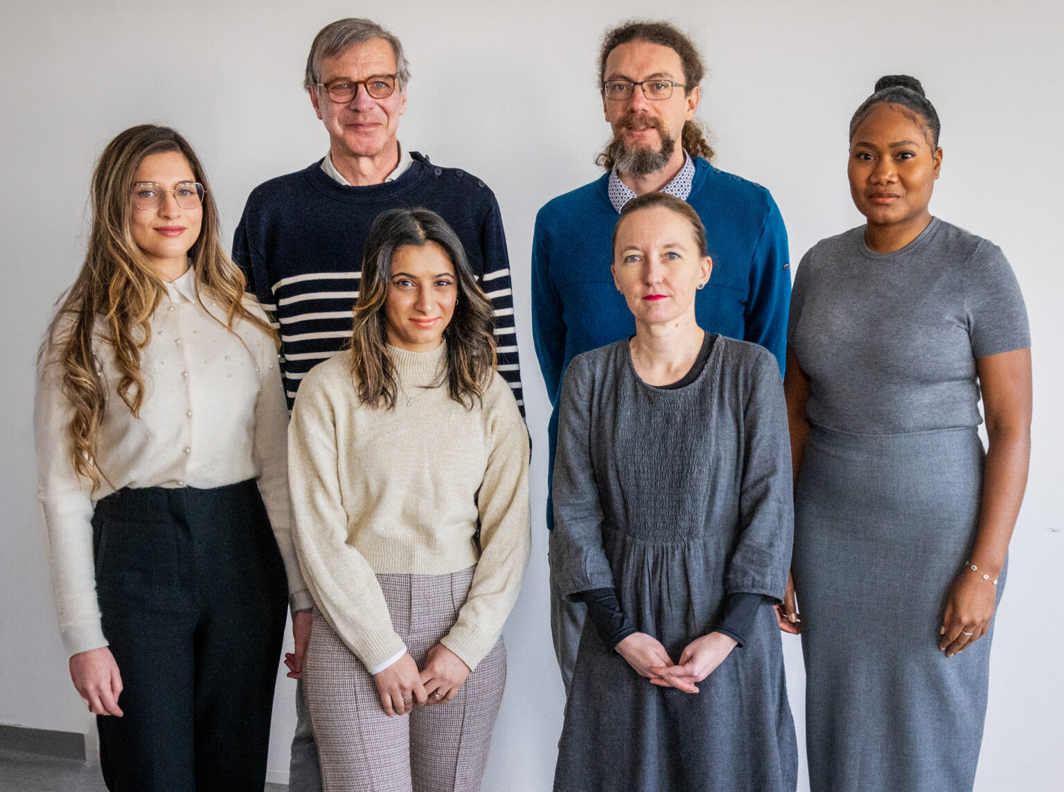
Biomarker Imaging Laboratory
Team Garteiser / Van Beers
Scientific areas : Pathophysiology of inflammatory & fibrotic diseases axis

Team composition
Clinicians
PhD students
Laboratory of Imaging Biomarkers, LBI
The team conducts translational research in medical imaging aimed at developing and validating new imaging biomarkers for inflammation, fibrosis, and cancer. This research extends from basic biomedical imaging to preclinical and clinical research concerning liver and abdominal pathologies.
Methodological research focuses on rapid, functional, and quantitative imaging using magnetic resonance imaging (diffusion MRI, perfusion MRI, elastography, susceptibility testing, relaxometry) and ultrasound, as well as the study of new tracers for cellular imaging.
These developments are first validated through experimental research in phantoms, tissue slices, and small animals, and then transferred to clinical imaging.
The laboratory is part of the “France Life Imaging” research infrastructure network, whose Paris Centre Hub is coordinated by Philippe Garteiser, as well as the “Imaging of Living Systems” program at the University of Paris. The LBI laboratory is one of the two constituent teams of the FRIM preclinical imaging platform (Federation of Research in Multimodal Imaging, UMS34).
Main Research Projects
Imaging of Liver Inflammation in Metabolic Diseases
Non-invasive assessment of inflammation and hepatocyte damage is an important objective in the diagnosis of non-alcoholic steatohepatitis (NASH). The hospital-university research project (“RHU QUID-NASH”) that we are conducting with other academic and industrial partners aims to evaluate new non-invasive diagnostic methods for NASH in murine models of steatohepatitis and in a cohort of 600 diabetic patients with hepatic steatosis. We are developing a virtual liver biopsy approach based on the integration of multi-omics data, including quantitative data from ultrafast ultrasound and multiparametric MRI, encompassing measurements of multi-frequency viscoelastic properties, R1 and R2 relaxation rates*, and lipid composition. Recently, we initiated an MRI study on the frequency dispersion of the water diffusion coefficient in NASH (ANR “Stedi-NASH”). This method allows for probing diffusion within liver tissue at precise spatial scales to assess hepatocyte size and characterize their cytoplasmic content.

Parametric maps of steatosis (PDFF, left) and liver stiffness (right) in two patients with simple steatosis (top, S1A1F1 histologically) and steatohepatitis with advanced fibrosis (bottom, S2A3F3 histologically). The patient with steatohepatitis has a higher liver stiffness (3 kPa) than the patient with simple steatosis (1.4 kPa).
Imaging of Forces in Liver Cancer
To provide advanced characterization of the mechanical properties of tumors, it is possible to measure apparent elasticity at different levels of pre-compression. We measure the biomechanical properties of tissues under compression using magnetic resonance elastography to evaluate solid and fluid pressures within tumors, two parameters influencing tumor aggressiveness and the efficacy of targeted therapies (European H2020 project “FORCE”).

Elasticity maps under different compression levels of a human hepatocellular carcinoma xenografted into mice, showing an increase in apparent elasticity under compression (Pagé G et al. J Magn Reson Imaging 2019, Pagé G et al. Cancers 2021)
Hepatic functional MRI with contrast agents
Pharmacokinetics of hepatobiliary agents by imaging
Hepatic MRI with injection of hepatobiliary contrast agents
iaires (Gd-EOB-DTPA, Primovist) and pharmacokinetic modeling allows us to evaluate hepatocyte uptake, biliary excretion and reflux to the sinusoids. These hepatobiliary agents cross hepatocyte membranes via transporters (OATPs, MRP2, and MRP3), creating concentration gradients between the extracellular compartment, the hepatocytes, and the biliary compartment.

Hepatocyte uptake